

Photobiology and Biotechnology: Controlled Optical Irradiation of Cells and Tissue
Photobiology examines how optical radiation – from UV-C to the near-infrared – interacts with biological material: with DNA, proteins, photosensitizers, photoreceptors and cell structures. Biotechnology puts this knowledge to work as a deliberate tool – to inactivate microorganisms, control cellular signalling pathways, carry out photodynamic treatment of tissue, or drive the light-mediated modification of biomolecules. The central technical challenge is that biological systems do not respond to electrical lamp power or a blanket exposure time, but to the spectral irradiance actually arriving at the site of action, the resulting dose, and the wavelength-dependent effectiveness of individual photons. Without traceable optical measurement, any biological observation remains only partially reproducible.
How is the market or the technology developing?
No single, uniformly defined market category exists for "photobiology and biotechnology", because the field cuts across pharma, medical technology, basic research and optical instrumentation. More reliable figures come from adjacent, clearly bounded sub-markets that can serve as indicators of the underlying technological dynamics.
The market for phototherapy equipment was estimated at roughly 382.3 million US dollars in 2022 and is projected to grow to about 521.1 million US dollars by 2030, a compound annual growth rate of around 4.0 percent (Grand View Research, 2024). The considerably larger but technically related market for UV disinfection equipment stood at about 4.1 billion US dollars in 2024 and is expected to reach roughly 7.5 billion US dollars by 2033, a CAGR of about 7.1 percent (Grand View Research, 2025). Within that market, UV lamps and in particular UV-LED modules are growing disproportionately fast, with industry-wide growth rates reported at times above 15 percent per year for LED-based sources (PRNewswire/Strategic Market Research, 2022).
Technically, this growth mainly reflects a shift in the light sources used: away from broadband mercury-vapour lamps and towards narrowband UV-LEDs and excimer sources. That shift raises the bar for photobiological characterization, because every new wavelength combination requires its own action spectrum, its own safety profile under IEC 62471, and its own dosimetry. Growing regulatory attention – for instance the 2022 and 2023 updates of IEC 62471-6 for UV lamp products and IEC 62471-7 for visible radiation – is a direct consequence of this proliferation of source types.
The shift from mercury lamps to UV-LEDs changes not only cost and efficiency, but also the methods available: because LED arrays provide several wavelengths simultaneously and independently controllable, action spectra can now be determined directly and experimentally by varying the excitation wavelength, rather than relying on a handful of fixed lamp lines. The establishment of far-UVC sources around 222 nm opens up applications beyond classical disinfection (see UV disinfection), because their shallow penetration into tissue and their distinct photochemistry – evidenced by markedly higher quantum yields for the photolysis of certain classes of biomolecule – enable new safety profiles and mechanisms of action (Jing et al., 2024, Water Research). On the regulatory side, the Minamata Convention is driving the global phase-out of mercury-containing lamps for general lighting by 2027; specialised UV sources for research and disinfection are so far addressed through separate exemptions, but are also under increasing pressure to adapt (Minamata Convention on Mercury – Exemptions). In parallel, IEC and CIE have updated their safety standards for lamps and lamp systems – IEC 62471-6:2022 for UV lamp products including UV-LEDs, and IEC 62471-7:2023 for sources emitting predominantly visible light – making photobiological risk classification of new LED and laser sources increasingly binding (IEC Webstore).
How does optical radiation act on biological systems?
Biological effects arise from the absorption of photons by specific molecules, the chromophores: nucleic acids absorb preferentially in the UV-C and UV-B range, aromatic amino acids and flavins in the UV-A and blue range, porphyrins and other photosensitizers often in the blue and red visible range. When a photon of suitable energy hits a chromophore, it can trigger an electronic transition that starts a photochemical reaction – DNA damage, singlet-oxygen generation, or a conformational change in a photoreceptor protein, for example. The probability of that conversion is described by the quantum yield. Because absorption spectra are wavelength-dependent, the photobiological effectiveness of a source is not determined by its total power, but by the product of spectral irradiance and a wavelength-dependent action spectrum that states the relative efficiency of each wavelength for a given biological endpoint (BIPM SI Brochure, Appendix 3).
Which technologies and light sources are used?
A wide range of source technologies is available for the controlled irradiation of biological samples, differing in bandwidth, wavelength range, thermal load on the sample, and ageing behaviour.
Low-pressure mercury lamps emit almost monochromatically at 253.7 nm and are traditionally used for UV-C applications in microbiology and disinfection. Medium- and high-pressure mercury lamps deliver a broadband line spectrum from UV-B to visible and serve, among other things, as a low-cost source for photostability testing. Xenon arc lamps simulate sunlight (D65/ID65) with a filtered, continuous spectrum and are the reference source in pharmaceutical photostability testing under ICH Q1B. Excimer lamps, for example based on krypton chloride, emit quasi-monochromatically – at 222 nm, for instance – and are gaining relevance beyond pure disinfection because of their shallower penetration depth into skin and eyes and their distinct photochemistry (Ma et al., 2023). UV and visible LEDs are narrowband, switch almost instantaneously, contain no mercury, and can be combined into arrays spanning several wavelengths, which greatly simplifies recording action spectra directly. Lasers and laser diodes deliver high power density in a very small area and are used above all where light must be guided by fibre optics to a defined location, such as in optogenetics or focused photodynamic treatment.
| Technology | Characteristics | Advantages / Limits | Typical application |
|---|---|---|---|
| Low-pressure mercury lamp | quasi-monochromatic, approx. 254 nm | High UV-C efficiency, well established, low cost. Mercury content, ageing, slow ignition | Microbiological inactivation |
| Medium-/high-pressure mercury lamp | broadband line spectrum, UV-B to visible | High total output. Strong heat generation, hard to filter | Photostability testing, UV curing |
| Xenon arc lamp (filtered) | continuous, daylight-like spectrum | Reproducible reference spectrum (D65/ID65). Calibration effort, pulsed operation | Photostability testing, sunscreen research |
| Excimer lamp (e.g. KrCl*, 222 nm) | quasi-monochromatic, short-wave | Shallow penetration into tissue, mercury-free. Limited power density, specialised optics required | Far-UVC research, near-surface inactivation |
| UV/visible LED | narrowband, multiple wavelengths combinable | Low thermal load, fast switching, long life. Lower single-unit output than discharge lamps | Action-spectrum determination, PDT, optogenetics |
| Laser / laser diode | monochromatic, high power density, fibre-couplable | Precise focusing, targeted light delivery. High cost, safety overhead | Optogenetics, focused PDT |
Which process quantities are decisive?
The photobiological effect of an exposure is not determined by a single quantity, but by the interplay of several physical quantities that must be recorded separately.
Irradiance (in W/m² or mW/cm²) describes the radiant power hitting a surface and determines how quickly a dose is reached. Dose, or radiant exposure (in J/m² or mJ/cm²), is the time integral of irradiance and is the quantity usually correlated with a biological effect such as a log reduction or an erythema. Spectral irradiance (W/(m²·nm)) is the base quantity whenever several wavelengths are to be compared, combined or weighted by an action spectrum – any desired weighted quantity can be derived from it, but not the reverse. Effective, action-spectrum-weighted irradiance accounts for the fact that not every watt of optical power is equally effective; only the fraction weighted by the relevant action spectrum counts. Photon flux (photons per second and area, often given as µmol·m⁻²·s⁻¹) matters in photobiotechnology because photochemical reactions are ultimately photon-limited, not energy-limited. Exposure duration and sample temperature must be recorded independently, since thermal effects can overlay photochemical effects without that being apparent from the dose alone.
What limits the process or causes errors?
A common mistake is equating electrical lamp power with optical dose at the site of action. Between the lamp and the target structure lie distance, optics, possibly a filter, the vessel wall, a lid, the culture medium and – at dense seeding – the cells themselves; each of these layers attenuates the radiation in a wavelength-dependent way. A measurement taken above the sample vessel therefore describes the source, not the actual exposure of the cell layer.
Equally common is the assumption that stating a time alone is sufficient documentation. Under the Bunsen–Roscoe reciprocity law, the product of irradiance and time is constant for purely photochemical processes; for biological systems, however, this reciprocity only holds within a limited intensity and time window. Repair mechanisms, oxygen resupply and mass transport operate on their own timescales, so the same dose delivered at high irradiance over a short time can produce a different outcome than the same dose at low irradiance over a long time (overview of reciprocity failure in photobiology; study on photobiological reciprocity limits). Anyone comparing series of experiments should therefore state irradiance, time and dose separately, rather than treating a shorter exposure at higher intensity as a neutral conversion.
A third, often underestimated point is confusing literature doses with directly transferable process values: an inactivation dose of, say, 10 mJ/cm² reported for a particular 222 nm excimer source (Ma et al., 2023) is valid for the measurement geometry and matrix described there – transferring it to a different sample thickness, a turbid medium, or a different wavelength is not permissible without a new measurement. Finally, it is often underestimated that thermal and photochemical effects can overlap: a high-power, broadband source measurably heats the sample, and a purely thermally triggered reaction can only be separated from the photochemical effect if both effects are captured in isolation (Höhn et al., 2015).
What influence do vessel material, culture medium and geometry have?
The spectral transmittance of sample vessels is strongly wavelength-dependent and, in practice, one of the largest yet least documented sources of error. Standard polystyrene lids and plates can attenuate UV-C and UV-B radiation by several orders of magnitude more than UV-A or visible light, so an experiment documented simply as "UV irradiation" may in reality have delivered very different sample doses depending on the vessel used. Culture media themselves contain UV- and blue-absorbing components – phenol red as a pH indicator, or riboflavin, for example – which, under the Beer–Lambert law, attenuate irradiance exponentially with increasing layer thickness; even a few millimetres of medium can meaningfully reduce the dose reaching the bottom of the culture. At dense cell seeding, an "inner-filter effect" adds to this, where cells near the surface receive a larger share of the dose than those lying deeper. In microplates, meniscus formation at the liquid surface further produces an inhomogeneous, position-dependent irradiance within the same well. Wherever one of these influencing factors is unknown, the actual irradiance should be measured at the intended sample location rather than in the free beam.
Photon energy, quantum yield and action spectra
Two quantities explain why an identical dose does not automatically produce an identical effect once the wavelength changes: photon energy and quantum yield.
Photon energy follows from
Ephoton = h · c / λ
with Planck's constant h = 6.626 × 10⁻³⁴ J·s, the speed of light c = 2.998 × 10⁸ m/s and the wavelength λ. Shorter wavelengths therefore carry more energy per photon.
The quantum yield Φ describes how many photochemical events occur per absorbed photon:
Φ = (number of photochemical events) / (number of absorbed photons)
Φ is not a constant of wavelength; it depends on the specific molecule, its environment and often on the excitation wavelength itself (IUPAC Gold Book). A precondition for applying either quantity meaningfully is that the radiation is actually absorbed – photons that are not absorbed do not contribute to the reaction, regardless of their energy. The practical consequence: two sources with identical irradiance but different wavelength typically produce different photobiological effects – both because the target molecule absorbs to a different degree, and because the quantum yield itself can be wavelength-dependent.
Worked example: comparing photon energy at 222 nm and 254 nm
Question: UV disinfection and photobiology research increasingly use excimer sources at 222 nm instead of conventional low-pressure mercury lamps at 254 nm. How does photon energy differ between the two wavelengths, and what does that mean for photochemical effectiveness?
Assumptions: monochromatic radiation at λ₁ = 222 nm and λ₂ = 254 nm; h = 6.626 × 10⁻³⁴ J·s; c = 2.998 × 10⁸ m/s.
Model: Ephoton = h · c / λ
Calculation:
E₁ = (6.626 × 10⁻³⁴ J·s × 2.998 × 10⁸ m/s) / 222 × 10⁻⁹ m ≈ 8.95 × 10⁻¹⁹ J
E₂ = (6.626 × 10⁻³⁴ J·s × 2.998 × 10⁸ m/s) / 254 × 10⁻⁹ m ≈ 7.82 × 10⁻¹⁹ J
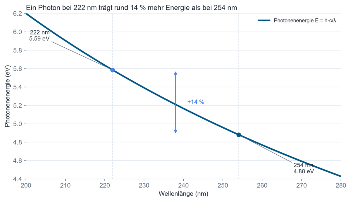
Converted to electronvolts (1 eV = 1.602 × 10⁻¹⁹ J): E₁ ≈ 5.59 eV, E₂ ≈ 4.88 eV.
Result: a photon at 222 nm carries roughly 14 percent more energy than a photon at 254 nm.
Technical interpretation: this difference corresponds with experimental findings that the quantum yield for photolysis of nucleobases and amino acids at 222 nm is 4 to 61 times higher than at 254 nm (Jing et al., 2024, Water Research). In practice, this means that an equal radiometric dose (J/cm²) at different wavelengths does not automatically produce a comparable biological effect – measuring and stating irradiance alone, without a wavelength reference, is not enough to describe a process reproducibly or to transfer it between sources.
Where is controlled optical irradiation used?
Dermatology and phototherapy. Narrowband UV-B (often around 311 nm) is used to treat psoriasis and vitiligo. The critical quantity here is the individual patient's minimal erythema dose (MED), which varies from person to person and is evaluated against the erythema action spectrum under ISO/CIE 17166.
Photodynamic therapy (PDT). A photosensitizer is excited by light whose wavelength is matched to its absorption bands – around 405 nm (Soret band) for superficial lesions, or 630–635 nm for deeper ones. The critical process quantity is penetration depth, which is greatest in the so-called therapeutic window between about 600 and 850 nm, where scattering and absorption by tissue are minimal (overview of PDT physics, PMC).
Cell and microbiology. In research laboratories, bacterial and cell cultures are irradiated under defined conditions to study DNA repair mechanisms, mutagenesis rates or phototoxicity, for example using a BS-02 irradiation chamber. The critical requirement is reproducibility of the dose at the actual sample location across many parallel runs and experiment days (Estévez Castro et al., 2018).
Pharmaceutical photostability testing. Under ICH Q1B, active substances and finished drug products must be exposed to a defined UV and visible-light dose (at least 200 W·h/m² UV and 1.2 million lux hours of visible light) to assess light-induced degradation. The critical factor is the exact spectral match of the test source to the D65/ID65 reference spectrum (EMA/ICH Q1B), covered in more depth under Pharma & Photostability.
Optogenetics and neuroscience. Genetically encoded photoreceptors such as channelrhodopsin (activated around 473 nm) or halorhodopsin (inhibited around 590 nm) are driven via fibre-coupled LEDs or lasers. The critical quantity is the irradiance at the fibre tip or at the target tissue, not the output power of the source (overview of optogenetic light sources, PMC).
Photobiotechnology and bioprocess engineering. Light is increasingly used as a selective tool in protein chemistry – for the light-driven functionalisation of biomolecules, for instance – as well as to illuminate photosynthetically active microorganisms in bioreactors (Berton & Holland, 2025). The critical factor here is photon flux per unit volume and the most homogeneous possible illumination of the reactor volume.
Cosmetics and sunscreen research. In-vitro tests of UV filter and photosensitizer efficacy use erythema- or phototoxicity-weighted irradiation. The critical factor is how closely the test spectrum matches the solar UV spectrum, since individual active-ingredient combinations can be strongly wavelength-selective.
How is irradiance measured at microplates, pulsed sources and fibre tips?
For highly parallel screening in microplates, LED arrays with individually addressable wavelengths and positions are used to compensate for spatial inhomogeneity by calibrating individual wells.
For pulsed sources such as xenon flash lamps, a purely time-integrating measurement is insufficient when the pulse shape itself is photobiologically relevant; time-resolved measurement methods are required here.
For thermally sensitive samples – live tissue samples or temperature-sensitive enzymes, for example – cooled sample holders with continuous temperature monitoring are combined to cleanly separate photochemical from thermal effects.
For in-vivo applications such as fibre-optic optogenetics or interstitial PDT, measurement in the free beam is fundamentally inadequate; irradiance must instead be estimated at the distal fibre tip or via a validated tissue model.
Which quantities must be measured or monitored?
The starting point for any measurement plan is which quantity actually matters for the biological endpoint in question – not which instrument happens to be available. If only a single, well-characterised wavelength is used and its action-spectrum weighting is already known, a broadband radiometer such as the RMD Pro with a filter combination matched to that action spectrum can be sufficient (see Selecting UV sensors for background on sensor choice). As soon as several wavelengths are to be compared, combined or newly evaluated, or when an unknown or broadband, ageing source is involved, a spectral measurement with a spectroradiometer such as the SR900 is required, because no spectral information can be recovered from an integral reading afterwards.
Measurements should always be taken at the actual sample location – at the position of the cell layer, not in the free beam above the vessel lid – because otherwise attenuation by the vessel material and medium is systematically underestimated. Relevant measurement uncertainties arise from spectral mismatch between sensor responsivity and the actual source spectrum, from the angular dependence of sensor responsivity (cosine error) under non-parallel incidence, and from the calibration uncertainty of the reference standard used. Spatially resolved measurements become necessary once a sample is larger than the uniformly illuminated area of the source – multiwell plates under a single LED array, for example. Temporal resolution becomes relevant for pulsed or drifting sources whose output changes within the exposure period.
In automated irradiation processes – series experiments with many samples over weeks or months, for example – a one-time setting of lamp power and exposure time is not enough, because the actual irradiance changes continuously through lamp ageing, contamination of optics, and temperature drift. Dose-controlled operation – as implemented by an UV-MAT irradiation chamber – continuously measures irradiance with a calibrated sensor and automatically ends the exposure once the target dose is reached, instead of switching off after a fixed time. That decouples the actually delivered dose from the ageing of the source and keeps series of experiments comparable over long periods.
For regulated environments – GMP-adjacent testing laboratories or accredited calibration laboratories under DIN EN ISO/IEC 17025:2018-03, for example – traceable, documented logging of every individual dose is additionally relevant, so batch or experiment comparisons remain verifiable afterwards; traceability of the sensors used is provided by the accredited calibration laboratory.
In practice, spectral irradiance is usually captured with a calibrated spectroradiometer covering UV, visible light and, where relevant, the near-infrared range in a single measurement. For routine measurements on known sources, radiometers with action-spectrum-weighted sensors are used, whose spectral responsivity has already been shaped to match a defined action spectrum. For defined, dose-controlled exposures of cell and bacterial cultures, irradiation chambers such as the UV-MAT with independently switchable spectral channels are used, whose dose control is based on the actually measured – not merely the nominal – irradiance.
What does published research show?
Experimentally separates the purely thermal from the photochemical effect of an exposure – exactly the kind of overlap that longer exposures often obscure.
Actual isothermal effects of water-filtered infrared A-irradiation
Höhn, Annika, et al. "Actual isothermal effects of water-filtered infrared A-irradiation." Photochemistry and Photobiology 91.4 (2015): 887–894.
Shows via gene-expression data that 222 nm irradiation directly photolyses repair enzymes, and supplies the markedly higher quantum yield for photolysis of nucleobases and amino acids at 222 nm versus 254 nm that underlies this page's worked example.
Suppression of photoreactivation of E. coli by excimer far-UV light (222 nm) via damage to multiple targets
Jing, Zi-Bo, et al. "Suppression of photoreactivation of E. coli by excimer far-UV light (222 nm) via damage to multiple targets." Water Research 255 (2024): 121533.
Characterises inactivation kinetics for several pathogens and surrogates under 222 nm excimer irradiation, and supplies the dose order of magnitude for far-UVC disinfection cited on this page.
UV Inactivation of Common Pathogens and Surrogates Under 222 nm Irradiation from KrCl* Excimer Lamps
Ma, Ben, et al. "UV Inactivation of Common Pathogens and Surrogates Under 222 nm Irradiation from KrCl* Excimer Lamps." Photochemistry and Photobiology (2023).
What does published research from customers show?
The following works by users show the breadth of the field – from skin reactions through repair mechanisms to light-driven protein chemistry.
Investigates how UV exposure changes the inflammatory response of skin immune cells.
Influence of UV irradiation on the skin-immune cell inflammatory response
Miceli, Rebecca, et al. "Influence of UV irradiation on the skin-immune cell inflammatory response." Research Square (2026).
Selects plant-derived compound mixtures for sunscreen formulation based on efficacy and cytotoxicity – treating protective effect as a spectrally weighted quantity.
Rational Selection of Phytochemical Mixtures for Sunscreen Development Based on in vitro Efficacy and Cytotoxicity Profiles
Martínez, Silvia Ximena Barrios, et al. "Rational Selection of Phytochemical Mixtures for Sunscreen Development Based on in vitro Efficacy and Cytotoxicity Profiles." Revista Brasileira de Farmacognosia 36.1 (2026).
Compares radiation and phototoxicity in the same cell lines and evaluates the enhancing effect of several photosensitizers, irradiated with a BS-04 irradiation chamber.
Comparison of Radio- and Phototoxicity in Association with an Enhancing Effect of the Photosensitizers Psoralen, Trioxsalen and Ortho-Iodo-Hoechst33258
Tietze, Katja, et al. "Comparison of Radio- and Phototoxicity in Association with an Enhancing Effect of the Photosensitizers Psoralen, Trioxsalen and Ortho-Iodo-Hoechst33258 on FaDu, PC-3, 4T1 and B16-F10 Cells." Biomedicines 13.1 (2024): 73.
Shows in repair-deficient mutants how strongly cellular repair determines the outcome of a UV exposure.
Influence of uvrA, recJ and recN gene mutations on nucleoid reorganization in UV-treated Escherichia coli cells
Estévez Castro, Carlos Felipe, Jorge Humberto Serment-Guerrero, and Jorge Luis Fuentes. "Influence of uvrA, recJ and recN gene mutations on nucleoid reorganization in UV-treated Escherichia coli cells." FEMS Microbiology Letters 365.11 (2018): fny110.
Uses light as a selective tool in protein chemistry rather than as a stress factor.
Light-induced chemistry for protein functionalisation
Berton, Cesare, and Jason P. Holland. "Light-induced chemistry for protein functionalisation." Chemical Communications (2025).
Across the photobiology, biotechnology and medicine topic areas, Opsytec customers have published 34 works in total; they are listed under Publications by customers – by topic.
Technical background and further sources
- ISO/CIE 17166:2019 – Erythema reference action spectrum and standard erythema dose (iso.org)
- IEC 62471 (series) – Photobiological safety of lamps and lamp systems, incl. IEC 62471-6:2022 (UV lamp products) and IEC 62471-7:2023 (visible radiation) (webstore.iec.ch)
- DIN 5031-10:2018 – Optical radiation physics and illuminating engineering, Part 10: Photobiologically effective radiation, quantities, symbols and effects (dinmedia.de)
- ICH Q1B – Photostability Testing of New Active Substances and Medicinal Products (ema.europa.eu)
- BIPM, SI Brochure, Appendix 3 – Units for photochemical and photobiological quantities (bipm.org)
- ISO 21348:2007 – Definitions of Solar Irradiance Spectral Categories (UV-A/-B/-C boundaries) (iso.org)
FAQ on photobiology and biotechnology
How does the biological effect of UV radiation work?
UV photons are absorbed by chromophores such as DNA bases or proteins and trigger photochemical reactions – DNA damage or structural changes, for example. Effectiveness depends strongly on wavelength, because every chromophore has its own absorption spectrum. Without absorption, no effect occurs, regardless of the power applied.
Which wavelength is suitable for photodynamic therapy?
The wavelength is chosen to match the absorption spectrum of the photosensitizer used. Blue light around 405 nm suits superficial lesions, red light around 630–635 nm suits deeper regions because it penetrates tissue more strongly. The choice is therefore a trade-off between absorption efficiency and penetration depth.
What dose is required for microbial inactivation?
The required dose depends on wavelength and organism. For 222 nm excimer sources, studies report doses around 10 mJ/cm² for a 4-log reduction of many bacteria and viruses; 254 nm sources often require comparable values, though with different effects on tissue. Literature values always apply to the specific measurement geometry and cannot be transferred without verification.
What is the difference between irradiance and dose?
Irradiance (W/m²) describes instantaneous power per unit area; dose (J/m²) is its time integral. It is typically the dose that correlates with a biological effect, while irradiance determines how quickly that dose is reached. Both quantities should therefore be documented separately. How irradiance, dose and fluence differ formally is set out in the overview of radiometric quantities.
Why does an identical dose at different wavelengths produce different effects?
Because photon energy and quantum yield are both wavelength-dependent. Shorter wavelengths carry more energy per photon and can activate different photochemical reaction pathways, leading to markedly different quantum yields. A dose figure alone, without a wavelength reference, is therefore insufficient for comparing effects.
How is the actual irradiance of a cell culture measured?
Ideally at the actual sample location, as close as possible to the cell layer, not in the free beam above the vessel. Vessel material, lid and culture medium attenuate radiation in a wavelength-dependent way, sometimes by orders of magnitude. A measurement taken outside these absorbers systematically underestimates the actual attenuation.
When is a broadband measurement sufficient, and when is a spectral measurement needed?
A broadband measurement with an action-spectrum-matched sensor is sufficient when a single, well-characterised wavelength is used. As soon as several wavelengths are to be compared, combined or newly evaluated, or the source is unknown or broadband, a spectral measurement with a spectroradiometer is required.
What role does temperature play in photobiological experiments?
High-power sources measurably heat the sample, and thermally triggered reactions can overlay or amplify the photochemical effect. A clean separation requires either a cooled sample holder or a parallel isothermal control measurement. Without temperature control, an observed effect cannot be reliably attributed to irradiation alone.
Related application fields
Photobiology and biotechnology border on several neighbouring fields: medical phototherapy applies the same photobiological principles to physician-prescribed treatment of patients, pharma photostability tests the light sensitivity of finished products rather than a biological effect, and occupational safety and photobiological safety addresses the same physical quantities for unintended rather than deliberate exposure. Also adjacent are UV disinfection as a specialised inactivation application, and Plants, Agriculture & Food Technology for the photobiological effect on plant material.
Author: Dr. Mark Paravia
Dr.-Ing. Mark Paravia is the managing director of Opsytec Dr. Gröbel GmbH in Ettlingen and heads the accredited calibration laboratory. Following his research on pulsed xenon excimer discharges at the Institute of Lighting Technology at KIT, his current focus is on optical radiation measurement technology. He is a recognized UV expert, vice-chair of the DIN Standards Committee FNL 7 “Optical Radiation,” and a member of the DVGW Project Group on UV Disinfection.
Unsure what dose is actually reaching a cell layer through a lid, medium and vessel wall?
Unsure what dose is actually reaching a cell layer through a lid, medium and vessel wall? A sound photobiological assessment starts with a measurement at the real sample location – not in the lamp's free beam. For defined, dose-controlled irradiation of cell and bacterial cultures, the UV-MAT irradiation chamber is well suited; for the spectral characterisation of unknown or combined sources, the SR900 spectroradiometer; and for routine monitoring of known sources, the RMD Pro radiometer. Calibration and traceability of the sensors used are provided by the accredited calibration laboratory under ISO/IEC 17025. Get in touch about your measurement task.