

Medical Phototherapy: UV Radiation, Mechanisms of Action and Dose Measurement
Medical phototherapy is the medically prescribed exposure of skin or tissue to ultraviolet or visible optical radiation of defined wavelength, irradiance and dose. It is used for, among other conditions, psoriasis, atopic dermatitis, vitiligo, actinic keratosis and neonatal hyperbilirubinemia. Unlike incidental UV exposure, the irradiation here is deliberate: it follows a physician-defined dose protocol designed to bring about a specific effect – such as apoptosis of inflammatory T cells or the photochemical breakdown of bilirubin – without crossing the threshold to erythema, burns or long-term damage. The central physical quantities are the spectral irradiance at the treatment site, the resulting cumulative dose over time, and the action spectrum, which describes how strongly individual wavelengths contribute to the biological effect. The technical challenge lies less in generating the radiation than in reproducibly quantifying it at the patient.
How is the market for medical phototherapy developing?
Reliable market figures for "medical phototherapy" are inconsistent because different studies draw different market boundaries – devices, treatments, regional segments or indications are not defined uniformly. This spread is itself a relevant finding: one report puts the global "Phototherapy Treatment Market" at USD 2.2 billion (2025), growing to USD 3.6 billion by 2035 (CAGR 5.2 %); psoriasis accounts for the largest indication share at 42.7 %, and narrowband UVB the largest technology share at 36.4 % (Future Market Insights, 2025). A narrower view limited to UV phototherapy devices alone puts the figure at around USD 800 million (2024), with a CAGR of 5.9 % through 2035 (WiseGuy Reports, 2025). For neonatal phototherapy in Europe, a considerably smaller sub-market of around USD 135 million (2024) with moderate growth (CAGR 4.4 %) is reported (MarketDataForecast, 2024).
More technically significant than the absolute market size are the shifts behind it: the move from mercury-vapour fluorescent tubes to LED sources, the growing spread of home-use devices for outpatient treatment, and a 2023 revision of the device standard IEC 60601-2-57 for non-laser light therapy equipment (IEC, 2023). Each of these developments raises the requirements for traceable dose control: LED sources age differently from fluorescent tubes, home-use devices escape routine oversight by trained staff, and a tightened device standard demands documented evidence of the irradiance actually reaching the patient – not merely the device's rated power.
Five developments determine how dose is applied and documented in phototherapy:
Transition from fluorescent tubes to UV LED systems. LED sources reach full output without warm-up time and age more evenly over their service life than fluorescent tubes, making time-based protocols more reliable over a longer period. At the same time, the narrower, steeper-edged emission tightens the requirement for a spectral sensor calibration matched to the specific LED – an effect less pronounced for broader fluorescent sources.
Growing importance of home-use devices. Market analyses report more than 46,000 portable phototherapy devices installed in the field for 2024 (Market Growth Reports, 2025); the technical consequence is a shift in quality assurance from clinical staff to factory-more-stable, low-maintenance light sources with built-in self-checking.
Revised device standard IEC 60601-2-57 (2023 edition). The second edition of the standard for non-laser light therapy equipment tightens the requirements for basic safety and performance evidence of therapeutic, diagnostic and cosmetic light sources, directly affecting the testing and documentation manufacturers must provide (IEC, 2023).
Growing adoption of daylight PDT. As a resource-efficient alternative to red-light PDT, it reduces the need for artificial light sources but in turn requires an assessment of the day's actual UV and light conditions, since solar irradiance cannot be controlled.
Inline spectral sensing in process and treatment systems. Miniaturised spectroradiometers such as the iSR900 enable continuous, rather than periodic manual, monitoring of spectral composition during operation, allowing earlier detection of spectral drift caused by ageing or contamination of optical components.
How does UV and light radiation act on the skin?
The therapeutic effect arises from the absorption of photons in skin or blood components and the resulting photochemical or photobiological reaction. In the UVB range (280–315 nm), the radiation is absorbed predominantly in the epidermis; DNA bases form cyclobutane pyrimidine dimers, triggering DNA repair processes, apoptosis of inflammatory T cells and modulation of cytokine signalling pathways – the postulated main mechanism behind narrowband UVB's efficacy in psoriasis (Ozawa et al., 1999). UVA (315–400 nm) penetrates deeper into the dermis and, combined with psoralens (PUVA), acts through DNA photoadducts and oxidative stress. In photodynamic therapy, a photosensitiser (e.g. protoporphyrin IX) is activated by visible light and, in the presence of oxygen, generates reactive oxygen species that selectively destroy damaged tissue. Neonatal bilirubin phototherapy uses blue light (450–460 nm) to photochemically convert unconjugated bilirubin into water-soluble isomers that can be excreted renally – not a DNA effect, but pure photoisomerisation.
Which technologies are used in phototherapy?
The light sources used differ in spectral width, coherence and penetration depth – properties that help determine the indication.
| Technology | Characteristics | Advantages / limitations | Typical use |
|---|---|---|---|
| Broadband UVB | 280–320 nm, fluorescent tube | Low cost, established. Contains erythemogenic short wavelengths with no added therapeutic benefit | Historical, increasingly displaced |
| Narrowband UVB (NB-UVB) | 311 ± 2 nm, fluorescent tube (TL01) or LED | Highest efficacy at a lower erythema threshold than broadband UVB. Steep action spectrum requires tight spectral control | Psoriasis, vitiligo, atopic dermatitis (standard therapy) |
| PUVA (psoralen + UVA) | 320–400 nm after oral/topical psoralen administration | Effective for thicker plaques, deeper penetration. Phototoxicity, added oral/topical medication, higher long-term risk | Chronic plaque psoriasis, palmoplantar dermatoses |
| UVA1 | 340–400 nm, high-output emitter | Reaches the mid/deep dermis, effective without psoralen. High irradiance required, large device footprint | Acute atopic dermatitis, scleroderma |
| Excimer lamp/laser 308 nm | Monochromatic, coherent for laser variants | Locally confined, high-dose irradiation sparing healthy skin. Economical only for small lesion areas | Vitiligo patches, treatment-resistant plaques |
| Blue-light phototherapy | 450–460 nm, LED or fluorescent tube | No DNA effect, favourable benefit-risk ratio. Effect depends strongly on irradiated area and irradiance | Neonatal hyperbilirubinemia |
| Photodynamic therapy (PDT) | 405–635 nm + photosensitiser | Tissue-selective, good cosmetic outcomes. Photosensitiser kinetics and oxygen supply are additional variables | Actinic keratosis, superficial basal cell carcinoma, acne |
The shift from fluorescent tubes to LEDs affects all UV and blue-light modalities alike, though to differing degrees: for already narrowband modalities (NB-UVB, excimer), the LED emission profile changes the spectrum only slightly; for broader sources such as UVA1 emitters, it shifts the spectral composition more substantially.
Which process quantities are decisive?
Spectral irradiance E(λ) in W/(m²·nm) describes how much radiant power per area and wavelength interval reaches the treatment site. It is the starting quantity for every further calculation, because the biological effect is wavelength-dependent.
Erythema-weighted (or effect-weighted) irradiance results from weighting E(λ) with a biological action function, for example the erythema reference action spectrum under ISO/CIE 17166. It is not a physical quantity but a biologically normalised one, and is therefore only meaningfully interpreted in the context of the specific action function it is based on.
Irradiation dose (radiant exposure) H, the product of irradiance and time, determines the cumulative photochemical effect. Treatment protocols fundamentally prescribe a dose, not a time – time is a derived control variable that can only be calculated correctly if the current irradiance is known.
Position and geometry determine which irradiance actually reaches the tissue. In whole-body cabinets it varies between head, torso and leg height and with body curvature; for handheld devices, it varies with the distance to the skin.
Exposure duration does not act purely additively through the dose; at very short or very long exposure times it can deviate from the simple dose-response relationship (see the reciprocity law).
What limits the process or causes errors?
A common misconception is to equate a lamp's electrical power with the optical dose delivered to the patient. A source's rated power describes the electrical power drawn, not the power actually emitted, and even less the radiant power reaching the treatment site. Between lamp and skin lie ballast efficiency, reflector geometry, distance, ageing and – in cabinets – shadowing from the body shape itself.
Ageing changes not only the magnitude but also the spectral distribution of the emission. In fluorescent tubes, irradiance drops disproportionately during the first operating hours, followed by a flatter, roughly linear decline; a time-based protocol without accompanying measurement therefore systematically overestimates the actual delivered dose as the tubes age. British clinical guidelines accordingly recommend re-measuring the irradiance used for dose calculation every 25–50 operating hours, and after just 10–15 hours following installation of new tubes, because fresh lamps initially age faster (Taylor et al., 2002). More on the underlying mechanisms is covered under UV lamp ageing.
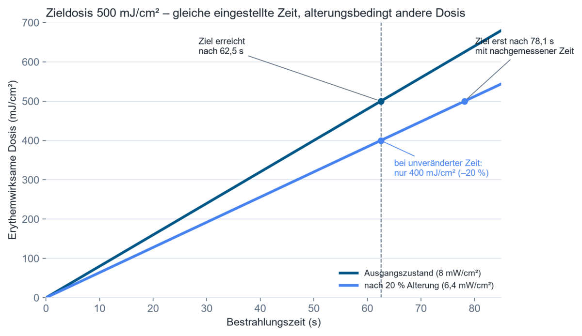
A time setting alone is valid only as long as the irradiance it is based on remains unchanged. If a cabinet's irradiance drops by 20 % through ageing, the time needed for the same dose increases by 25 % – if this is not re-measured, the patient permanently receives too low a dose at the same set exposure time.
Homogeneity is routinely underestimated. In whole-body cabinets, irradiance is typically highest at the torso and falls off towards the head and knees; a fixed body posture creates "shielded zones" with markedly lower dose. Clinical protocols therefore call for multi-point measurements – for example four positions each at chest, waist and knee height – whose mean value serves as the irradiance reference for dose calculation (Taylor et al., 2002).
The measuring instrument's spectral response must match the source. In narrowband UVB, the erythema-effective weighting changes by several orders of magnitude between 300 and 320 nm; if a source's emission peak shifts by only a few nanometres, the effective dose changes substantially, while a broadband instrument barely registers a change. A sensor calibrated for broadband sources can therefore deviate systematically at a narrowband source – this effect (spectral mismatch) is described, among others, in CIE 220:2016 and covered in more depth under Spectral mismatch for UV sensors.
Reciprocity does not hold unconditionally. The Bunsen-Roscoe law states that the photochemical effect depends only on the product of irradiance and time, regardless of their ratio. For the exposure times typical in clinical practice (seconds to a few minutes), this assumption is largely reliable for the established action functions; however, it can fail for extremely short, high-intensity pulses (e.g. pulsed excimer sources) or very long, low-dose exposures, because cellular repair mechanisms or saturation effects then compete with the primary photochemical reaction.
Action spectrum and dose quantities: the technical background
The photobiological effectiveness of an exposure does not follow from the unweighted irradiance, but from its convolution with an action function s(λ). For erythema, ISO/CIE 17166:2019 defines the erythema-effective irradiance as:
Eer = ∫ E(λ) · ser(λ) dλ (integrated over 250–400 nm)
where E(λ) is the source's spectral irradiance and ser(λ) the CIE reference erythema action spectrum. The resulting erythema-effective dose Her = Eer · t is often expressed in Standard Erythema Dose (SED); by definition, 1 SED equals 100 J/m² of erythema-effective exposure (ISO/CIE 17166:2019). The same logic – an action function multiplied by the spectrum, integrated over time – also underlies the bilirubin-effective irradiance used in neonatology, though there without a steep weighting function, simply as an integral over 400–550 nm.
Photon energy offers a complementary, often underappreciated perspective: Ephoton = h · c / λ. At 311 nm (typical NB-UVB central wavelength), it amounts to roughly 4.0 eV (6.4 × 10⁻¹⁹ J) – enough to affect covalent bonds in DNA, but too low in energy to penetrate much beyond the upper skin layers. At 450 nm (blue-light phototherapy), photon energy drops to about 2.75 eV; there, the dominant process is not DNA damage but the photochemical isomerisation of the bilirubin molecule. Wavelength therefore determines not only penetration depth but the mechanism of action itself.
Practical consequence: two devices with identical unweighted irradiance can differ in efficacy if their spectra differ in how strongly they populate the effective wavelength range. A broadband measurement in W/cm² is therefore sufficient only when the spectrum and the action function are known to match well; otherwise a spectral measurement is required.
Worked example: calculating treatment time for an NB-UVB session
Assumptions: a whole-body cabinet with narrowband UVB tubes (311 nm); target dose under a common dosing protocol for Fitzpatrick skin types III–IV: 500 mJ/cm² (AAD-NPF protocol, cited via cmsderm.ca, 2026); measured, averaged irradiance at the patient (designated patient irradiance) at the start of treatment: 8 mW/cm² (within the typical 6–8 mW/cm² range for NB-UVB cabinets).
Model: time t = dose H / irradiance E.
Calculation (initial state):
t = 500 mJ/cm² ÷ 8 mW/cm² = 62.5 s.
Calculation (after 20 % ageing of the tubes):
E' = 8 mW/cm² × 0.8 = 6.4 mW/cm²
t' = 500 mJ/cm² ÷ 6.4 mW/cm² = 78.1 s.
Technical interpretation: if the time set on the device remains at 62.5 s even though irradiance has dropped to 6.4 mW/cm², the patient receives only 400 mJ/cm² instead of the prescribed 500 mJ/cm² – a 20 % underdose, with no error visible on the device itself. Accumulated over a multi-week treatment cycle, this affects the response rate. The only way to detect it is regular, spatially resolved re-measurement of the actual irradiance – not reliance on a value determined once at the start, for example with the RMD Pro radiometer.
Where is medical phototherapy used?
Dermatology (psoriasis, atopic dermatitis, vitiligo). Narrowband UVB is the most commonly used modality here; the critical process quantity is precise adherence to the individual, skin-type-dependent starting dose and its stepwise escalation, because the therapeutic window between effective and erythema-inducing dose is narrow.
Neonatology (hyperbilirubinemia). Blue-light phototherapy in incubators or warming beds requires irradiance, measured spectrally in the 460–490 nm range, of typically ≥ 30 µW/(cm²·nm) across the entire treated skin area for "intensive phototherapy" (American Academy of Pediatrics, 2022; Dam-Vervloet et al., 2021). What matters here is less the peak irradiance at the centre than the size of the area over which this threshold is still reached – the so-called irradiance footprint.
Dermatologic oncology and aesthetic medicine (PDT). In photodynamic therapy, besides light dose and wavelength, the photosensitiser concentration in the target tissue and local oxygen availability are critical; insufficient exposure area or homogeneity leads to incomplete cell destruction and recurrence risk.
Research and photobiology. In cell and tissue models, UV or light irradiation is applied under controlled conditions that are usually far more tightly defined than in the clinic, for example using a BS-02 irradiation chamber; here, exact reproducibility of exposure conditions between experiments takes precedence over adapting to individual patient anatomy.
What are home devices, excimer handpieces, daylight PDT and light blankets suited for?
Home phototherapy devices allow treatment without repeated clinic visits and thereby improve treatment adherence, but shift more of the responsibility for correct use and device oversight onto the patient – a loss of control that is partly offset by factory-more-stable, usually LED-based light sources and automated timing.
Excimer handheld devices (308 nm) allow locally high-dose treatment of individual lesions while sparing surrounding healthy skin; their small treatment area makes them uneconomical for widespread disease but well suited to circumscribed lesions such as vitiligo patches or treatment-resistant plaques.
Daylight-mediated PDT uses natural sunlight instead of an artificial light source to activate the photosensitiser in actinic keratosis; it reduces pain compared with conventional red-light PDT, but depends on weather conditions and UV index and is therefore harder to standardise (European Dermatology Forum, 2019).
Fibre-optic phototherapy systems in neonatology (e.g. light blankets) allow irradiation without separating mother and child, but present smaller and geometrically different irradiance footprints than overhead emitters, which must be accounted for in dose planning.
Which quantities must be measured or monitored?
The starting point for any measurement plan is the question of which quantity the treatment protocol actually requires – generally the irradiance at the skin surface, not at the lamp housing. A broadband radiometer such as the RMD Pro, with a spectral response matched to the action function, is sufficient as long as the calibration source and the actual treatment source are spectrally similar enough (see how UV sensors work). Where the emission spectra of several device types in use differ, or where the spectral composition itself needs to be checked – for example when comparing a new LED source with an existing fluorescent-tube cabinet – a spectral measurement with a spectroradiometer such as the SR900 is required, because any desired action function can then be derived computationally.
Spatially resolved measurements become necessary once the irradiated area is larger than the sensor and homogeneity is unknown – for example in whole-body cabinets or neonatal irradiation units with an undefined footprint. Time-resolved measurements are relevant when a source is unstable (warm-up behaviour, mains fluctuations) or when checking whether the reciprocity assumption still holds for the planned exposure duration.
Many treatment cabinets and incubators have built-in sensors that measure irradiance in real time and automatically calculate the exposure time from the target dose and the current reading. These built-in dosimeters, however, typically capture only reflected light from a limited area of skin and thus only approximate the average irradiance across the whole patient; if the internal reading deviates by more than about 10 % from a direct external measurement, clinical guidelines recommend readjustment or switching to time-based operation with externally validated values (Taylor et al., 2002).
For regulated medical operation, documentation requirements are also relevant: every treatment session – dose, duration, wavelength used, device status – must be traceably recorded to ensure traceability of effect and any side effects under medical device regulation. The technical significance lies not in the digital interface itself, but in the fact that only an unbroken link between measured value, timestamp and device calibration status makes it possible, after the fact, to distinguish an under- or overdose from a lack of efficacy or an unexpected side effect.
In clinical practice, an achievable measurement uncertainty of around 10 % is generally considered sufficient; geometric effects (patient position, body curvature) can push this uncertainty to 15 % or more in practice (Diffey/Hart, cited via the Dundee guidelines). Traceability of the sensor is not a formality but a precondition, because the measured value directly determines the treatment time and therefore the dose actually delivered to the patient – ensured via the accredited calibration laboratory under DIN EN ISO/IEC 17025:2018-03, with fixed calibration intervals.
What does the published research show?
The T-cell apoptosis mechanism of narrowband UVB in psoriatic lesions was first systematically described in a bilateral comparison study documenting the difference in effect between broadband and 312 nm narrowband UVB on epidermal and dermal T cells:
Ozawa, M.; Ferenczi, K.; Kikuchi, T.; Cardinale, I.; Austin, L. M.; Coven, T. R.; Burack, L. H.; Krueger, J. G.: "312-nanometer Ultraviolet B Light (Narrow-Band UVB) Induces Apoptosis of T Cells within Psoriatic Lesions". Journal of Experimental Medicine, 1999, 189(4), 711–718. DOI: 10.1084/jem.189.4.711.
A recent systematic review assesses the efficacy of narrowband UVB for psoriasis specifically in patients with darker skin pigmentation, a field with previously underrepresented study data:
Hauptman, M.; El Othmani, A.; Pazhyanur, S.; Nakamura, M.: "Narrowband-Ultraviolet B Phototherapy for Psoriasis Treatment in Skin of Color: A Systematic Review and Meta-Analysis". Photodermatology, Photoimmunology & Photomedicine, 2025, 41(5), e70051. DOI: 10.1111/phpp.70051.
A review article summarises the pharmacological and photochemical basis of PUVA therapy and its current clinical role, which newer modalities such as NB-UVB have partly displaced:
Richard, E. G.: "The Science and (Lost) Art of Psoralen Plus UVA Phototherapy". Dermatologic Clinics, 2020, 38(1), 11–23. DOI: 10.1016/j.det.2019.08.002.
A report from a British professional society provides the methodological basis for dosimetry and calibration of UV therapy devices, including concrete measurement protocols for whole-body cabinets:
Taylor, D. K.; Anstey, A. V.; Coleman, A. J.; Diffey, B. L.; Farr, P. M.; Ferguson, J.; Ibbotson, S.; Langmack, K.; Lloyd, J. J.; McCann, P.; Martin, J. C.; Menagé, H. du P.; Moseley, H.; Murphy, G.; Pye, S. D.; Rhodes, L. E.; Rogers, S.: "Guidelines for dosimetry and calibration in ultraviolet radiation therapy: a report of a British Photodermatology Group workshop". British Journal of Dermatology, 2002, 146(5), 755–763. DOI: 10.1046/j.1365-2133.2002.04740.x.
A comparative study of six commercial neonatal phototherapy devices was the first to systematically quantify the "irradiance footprint" – the area over which the clinically relevant threshold irradiance is actually reached:
Dam-Vervloet, A. J.; Bosschaart, N.; van Straaten, H. L. M.; Poot, L.; Hulzebos, C. V.: "Irradiance footprint of phototherapy devices: a comparative study". Pediatric Research, 2021, 92(2), 453–458. DOI: 10.1038/s41390-021-01795-x.
A recent review places photodynamic therapy in mechanistic and clinical context, summarising which combination of photosensitiser, light dose and wavelength is documented for which dermatologic indication:
Di Guardo, A. et al.: "Photodynamic Therapy in Dermatology: A Comprehensive Review". International Journal of Molecular Sciences, 2026, 27(9), 3960. DOI: 10.3390/ijms27093960.
What does published research from customers show?
The following customer publications show the breadth of the field – from extracorporeal cell therapy through photodynamic oncology to comparative photosensitiser research methodologically related to PUVA therapy.
Investigates which immune cells mediate the effect of extracorporeal photopheresis – an established UVA- and psoralen-based phototherapy used to treat graft-versus-host disease and cutaneous T-cell lymphoma, carried out with a BS-02 irradiation chamber.
Suppressive CD8+ T-Cells Are Key Cellular Mediators of Extracorporeal Photopheresis
Rogers, Kai J., et al. "Suppressive CD8+ T-Cells Are Key Cellular Mediators of Extracorporeal Photopheresis." Journal of Clinical Apheresis 41.1 (2026).
Tests halogen-substituted photosensitisers for the photodynamic therapy of prostate cancer in vitro, irradiated with a BS-04 irradiation chamber.
Halogen substituted 4-thio-2′-deoxyuridines as photosensitizers for the photodynamic therapy of prostate cancer. An in vitro study
Czaja, Anna, et al. "Halogen substituted 4-thio-2′-deoxyuridines as photosensitizers for the photodynamic therapy of prostate cancer. An in vitro study." Spectrochimica Acta Part A: Molecular and Biomolecular Spectroscopy (2025): 126802.
Compares radio- and phototoxicity on the same cell lines and assesses the enhancing effect of the PUVA photosensitisers psoralen and trioxsalen – the same class of substance used in clinical PUVA therapy, irradiated with a BS-04 irradiation chamber.
Comparison of Radio- and Phototoxicity in Association with an Enhancing Effect of the Photosensitizers Psoralen, Trioxsalen and Ortho-Iodo-Hoechst33258
Tietze, Katja, et al. "Comparison of Radio- and Phototoxicity in Association with an Enhancing Effect of the Photosensitizers Psoralen, Trioxsalen and Ortho-Iodo-Hoechst33258 on FaDu, PC-3, 4T1 and B16-F10 Cells." Biomedicines 13.1 (2024): 73.
Further work from adjacent fields of photobiology is listed under Publications by customers, by topic.
Technical background and further sources
- ISO/CIE 17166:2019 – Erythema reference action spectrum and standard erythema dose (iso.org)
- IEC 60601-2-57:2023 – Medical electrical equipment, Part 2-57: Particular requirements for non-laser light source equipment for therapeutic, diagnostic, monitoring, cosmetic and aesthetic use (webstore.iec.ch)
- IEC 62471 – Photobiological safety of lamps and lamp systems (iec.ch)
- ICNIRP Guidelines on Limits of Exposure to Ultraviolet Radiation of Wavelengths between 180 nm and 400 nm, 2004 (icnirp.org)
- American Academy of Pediatrics: Clinical Practice Guideline Revision – Management of Hyperbilirubinemia in the Newborn Infant, 2022 (PDF)
- CIE 220:2016 – Characterization and Calibration Method of UV Radiometers
FAQ on medical phototherapy
How does medical phototherapy work?
It uses UV or visible light of a defined wavelength to trigger a specific photochemical reaction in skin or blood – for example, DNA-mediated apoptosis of inflammatory cells with narrowband UVB, or the photochemical conversion of bilirubin in neonatal blue-light therapy. Efficacy and safety depend on wavelength, irradiance and dose, not on exposure duration alone.
Which wavelength is used for psoriasis?
Narrowband UVB with an emission peak around 311 nm is considered the most effective UV modality, with a lower erythema risk than broadband UVB, because the steep action spectrum in this range favours the ratio of therapeutic effect to erythema risk. For localised, treatment-resistant lesions, the 308 nm excimer laser or excimer lamp is also considered, while PUVA remains relevant for thicker, more widespread plaques.
What UV dose is needed for a phototherapy session?
This depends on skin type and indication. Typical starting doses for narrowband UVB range from about 100 to 800 mJ/cm², depending on Fitzpatrick skin type and the protocol used, with stepwise escalation over the course of treatment. The individually tolerated dose is usually determined via a test irradiation (minimal erythema dose testing) or based on skin type.
What is the difference between PUVA and narrowband UVB?
PUVA combines UVA light (320–400 nm) with a photosensitising psoralen and acts through DNA photoadducts; narrowband UVB (311 nm) requires no additional substance and acts primarily through direct UVB-DNA effects and apoptosis induction. Narrowband UVB is today generally regarded as the first-line modality, while PUVA remains relevant for thicker, treatment-resistant plaques.
Why doesn't the same electrical lamp power always deliver the same treatment dose?
Electrical power describes the device's energy consumption, not the optical radiation reaching the patient. Ballast efficiency, reflector geometry, distance and, above all, ageing of the light source change the actual irradiance independently of rated power. Only a regular measurement at the treatment site shows whether the set time still corresponds to the prescribed dose. The underlying definitions and units are listed in the overview of radiometric quantities.
How is UV irradiance measured in a treatment cabinet?
With a calibrated radiometer whose spectral response matches the cabinet's emission spectrum, at several positions at head, torso and knee height. The mean of these measurements serves as the reference value for calculating treatment time; where several different source types are in use, an additional spectral measurement is advisable.
What factors, besides dose, affect the efficacy of a phototherapy session?
Relevant factors include the homogeneity of irradiation across the treatment area, individual skin pigmentation and photosensitivity, and – for PDT – the photosensitiser concentration and tissue oxygen supply, as well as, for neonatal phototherapy, the size of the adequately irradiated area (irradiance footprint). The spectral match between the measuring instrument and the light source also affects how reliably the actually effective dose is captured.
Related application fields
Medical phototherapy borders on several neighbouring fields: Pharma & Photostability tests the light sensitivity of finished products rather than irradiating people therapeutically, Photobiology & Biotechnology studies the effect of radiation on living systems in research, and occupational safety and photobiological safety cover the same physical quantities for unintentional rather than medically prescribed exposure (see also UV erythema in the workplace). The measurement side – dose control and GMP records – is covered by UV measurement in medicine and GMP settings, and standards and limit values under Guidelines, norms and standards in UV.
Author: Dr. Mark Paravia
Dr.-Ing. Mark Paravia is the managing director of Opsytec Dr. Gröbel GmbH in Ettlingen and heads the accredited calibration laboratory. Following his research on pulsed xenon excimer discharges at the Institute of Lighting Technology at KIT, his current focus is on optical radiation measurement technology. He is a recognized UV expert, vice-chair of the DIN Standards Committee FNL 7 “Optical Radiation,” and a member of the DVGW Project Group on UV Disinfection.
Unsure whether the correct dose is reaching the patient?
Unsure whether the set exposure time still matches the prescribed dose once the light source has aged since its last calibration? A spatially resolved, spectrally matched irradiance measurement at the treatment site provides clarity – regardless of which device is used to carry it out. For ongoing irradiance and dose monitoring at treatment devices, the RMD Pro radiometer is well suited; for the spectral characterisation and comparison of different source types, the SR900 spectroradiometer; and for defined, dose-controlled irradiation of cell or tissue samples in photobiological research, the BS-02 irradiation chamber. Calibration and traceability of the sensors used is ensured by the accredited calibration laboratory under ISO/IEC 17025. Get in touch about your measurement task.